


Oxford chemists capture the first “molecular movie” of penicillin biosynthesis
9 October 2026
- Chemists from the University of Oxford have captured the first atomic-resolution “molecular movie” that shows how nature builds the core structure of penicillin.
- The study solves a many decades-old mystery: how does nature construct the remarkable ring-shaped structure common to all penicillin and cephalosporin antibiotics?
- The findings will help guide the engineering of new classes of antibiotics, and the development of artificial enzymes, to tackle the growing challenge of antimicrobial resistance.
More than 80 years after Oxford scientists transformed medicine by developing penicillin into the world's first widely used antibiotic, researchers at the University of Oxford have revealed, for the first time, exactly how nature constructs the core structure of the drug.
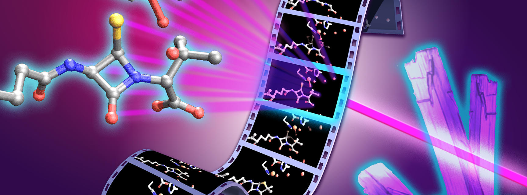
Published today in Nature Catalysis, the researchers captured penicillin biosynthesis in real time. The resulting atomic-resolution "molecular movie" reveals, step by step, how the enzyme IPNS (isopenicillin N synthase) builds the distinctive four-membered β-lactam ring that gives penicillin and related antibiotics their bacteria-killing activity.
Oxford has played a central role in the history of penicillin since the pioneering work of Nobel prize-winning scientists Prof Howard Florey and Sir Ernst Chain, and colleagues, who developed penicillin into the first clinically useful antibiotic during the Second World War. This new study ultimately builds on the work of Nobel prize-winning Oxford chemist Dorothy Hodgkin, who first solved the structure of penicillin in 1945 using X-ray crystallography.
Although scientists have studied the penicillin-forming reaction for decades, the fleeting intermediates involved had proved too short-lived to observe directly, and the complex ring-forming reaction has never been reproduced using conventional chemical catalysts.
The new study shows, in unprecedented detail, how the enzyme IPNS achieves an exceptionally complex transformation in a single step. This resolves a long-standing mechanistic question that has remained unanswered for more than four decades.
However, rather than relying on static X-ray crystallographic structures of the enzyme in its resting state, the researchers followed the reaction in real time using ultrafast X-ray free-electron laser (XFEL) experiments.
Penicillins, and related β-lactam antibiotics such as cephalosporins, account for more than half of the antibiotic doses administered worldwide each year. With rising rates of antimicrobial resistance – a process in which bacteria and other microorganisms evolve to survive the medicines designed to kill them –understanding how nature performs this antibiotic-forming reaction is an important step towards replenishing the pipeline of new antibiotic drugs.
Dr Patrick Rabe, Wellcome Mid Career Development Award Investigator in the Department of Chemistry and lead author of the study, said:
These insights give us a much richer understanding of how IPNS controls a remarkably difficult chemical transformation. The ability to capture structural snapshots over milliseconds to seconds of reaction time at a macromolecular level allows us to connect enzyme motion, iron chemistry and water-mediated proton transfer. By understanding this process in atomic detail, we are uncovering design principles that nature uses to control highly reactive chemistry. These principles could help guide the engineering of enzymes for applications ranging from antibiotic discovery to sustainable catalysis.
Professor Christopher Schofield, Professor of Chemistry at the University of Oxford and a senior author of the study, said:
Penicillin has shaped modern medicine, but there was still much to learn about how nature builds this important antibiotic structure. By capturing the structure of fleeting intermediates, we can better understand how enzymes control highly reactive intermediates with remarkable precision. The results reveal the importance of water molecules in controlling the penicillin ring-forming reactions, something we are now exploring in the many human enzymes related to IPNS.
To capture the reaction in real time, the researchers used a system in which thousands of tiny droplets containing anaerobic enzyme microcrystals were deposited onto a moving 2 mm wide tape. As the tape entered an oxygen filled chamber, oxygen rapidly diffused into the crystals and initiated the reaction simultaneously across the sample. By precisely controlling the speed of the tape, the researchers could determine how long each crystal reacted before reaching the X-ray interaction point, where an ultrafast XFEL pulse recorded an atomic resolution snapshot.
Combining thousands of these snapshots allowed the team to build a frame-by-frame “molecular movie” of penicillin biosynthesis. This method allows individual reaction intermediates that exist only for tiny fractions of a second to be observed at atomic resolution, and under physiological temperature and pressure, before they disappear.
The researchers discovered several previously unknown reaction intermediates, including the first observation of a monocyclic β-lactam intermediate. They also identified a previously unseen thioaldehyde intermediate that transiently dissociates from the catalytic iron centre before re-coordinating to the metal centre after formation of the monocyclic β-lactam ring. Together, these findings reveal an unexpected level of control over the reaction. They also found that water molecules inside the enzyme actively direct the chemistry and that structural changes throughout the protein – not just at the catalytic iron centre – drive the reaction.
The study combined time-resolved XFEL crystallography with spectroscopy organic synthesis, computational chemistry and biochemical experiments to reveal how the enzyme builds penicillin at atomic resolution. The work brought together researchers in chemistry, biology, physics, engineering and computational science, with experiments carried out at major international X-ray facilities in the UK, USA, Japan and the Republic of Korea.
Beyond penicillin, IPNS belongs to a large family of iron-dependent oxygenase enzymes involved in human biology, including ones that enable us to sense and respond to changes in oxygen availability. The mechanistic insights from this work therefore have implications far beyond antibiotics, offering new principles for enzyme engineering and catalyst design.
Dr Allen M Orville, group leader of the XFEL Hub and co-author of the paper, said:
The XFEL Hub at Diamond brought specialist expertise in time-resolved X-ray methods and XFEL-based structural biology to this study. Working closely with our colleagues in Oxford and at partner facilities, we helped bring together the experimental approaches needed to capture these fleeting stages of enzyme catalysis. The results show the power of combining complementary expertise and X-ray techniques to move beyond static structures and reveal how enzymes work in real time. It is a great example of how time-resolved structural biology can uncover new principles of enzyme function and, ultimately, inform the design of new catalysts and therapeutics.
You can read the full study in Nature Catalysis.
Image credits. Header image: University of Oxford/Greg Stewart/SLAC National Accelerator Laboratory.

