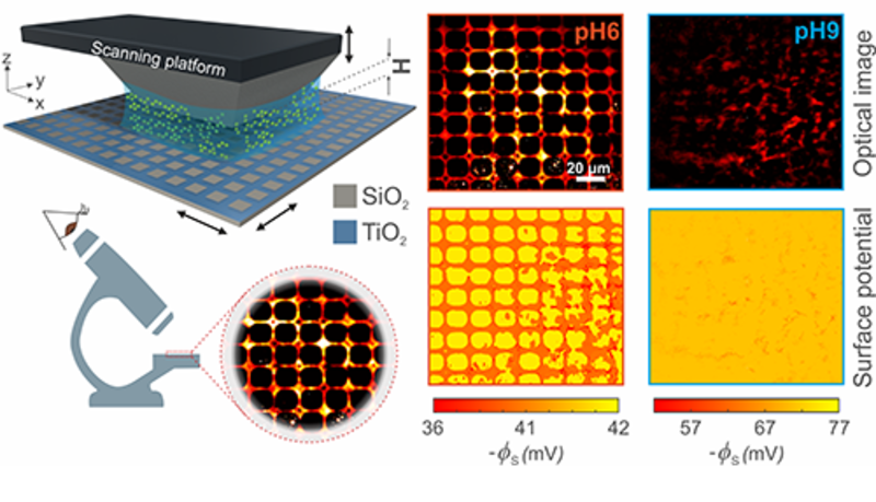
A new optical imaging technique developed in the Department of Chemistry will enable easy characterisation of the electrical properties of solid-liquid interfaces.
Surfaces immersed in liquids develop a net electrical charge that can have a profound impact on their properties and performance. This can be vital in any interface-governed context, from biofouling to energy production and storage devices. Currently there are few, if any, broadly applicable techniques that permit interfaces and their spatial chemical variation to be characterised and investigated in a straightforward way.

Optical imaging of the electrical charge on a glass substrate coated with with a thin film of titanium dioxide in a grid pattern. Regions of bright intensity indicate that when the pH in solution is low, titanium dioxide has a lower electrical potential compared to glass (silicon dioxide) which appears much darker in the image.
In a study recently published in the Proceedings of the National Academy of Sciences, a team from Professor Madhavi Krishnan’s research group used simple wide-field fluorescence microscopy to optically image the electrical repulsion between a probe molecule and a test surface, thereby shedding light on the charge and chemical properties of the interface.
The technique supports monitoring of variations in surface charge on a millisecond timescale and over millimetre length scales and will help researchers better characterise and understand interfaces. Future work will look to take the method beyond the measurement of electrical charge and apply the principle to the investigation of others types of forces between molecules and surfaces.
Professor Krishnan says:
“In the course of our daily activities in the lab, we would constantly find ourselves in need of knowing the amount of electrical charge carried by various types of surface-materials and coatings such as inorganic oxides or polymers. We were invariably stumped for an answer, as there were no simple and generally applicable techniques that could provide a quick and reliable read-out.
“We have now developed a technique where a charged light-emitting molecule, driven around in solution for free by thermal energy, acts as a mobile probe of the forces due to nearby surfaces. Optical observation of a number of such thermally mobilised probe molecules in parallel permits us to read out the properties of the surface, in much the same way that an AFM (atomic force microscopy) tip moving over a surface mechanically responds to the force it experiences.
“It also turns out that our approach is about a million times faster than a cantilever-based mechanical method. I am very excited about this development in our lab, and want to applaud the efforts of my graduate student Sushanta Mahanta who has been instrumental in realising the project.”
You can read more about the study here.